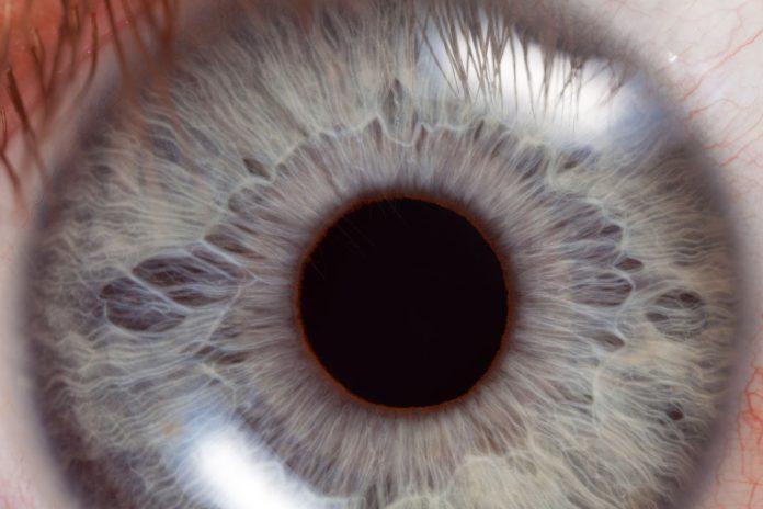
Neurological exams are crucial in diagnosing and monitoring brain health, providing invaluable insights into a patient’s cognitive and motor functions. One critical component of these exams is the pupil reactivity test, which assesses how a patient’s pupils respond to light.
This article will delve into the importance of precise pupil diameter measurement, discussing its implications for neuro exams, the factors that affect it, and the advanced neurological tools and techniques available for enhanced assessment. By understanding the significance of pupillary size measurement and embracing innovative methods, health professionals can improve the overall quality of neurological examinations.
Background on Neurological Exams
In clinical settings, neurological exams are vital for identifying potential issues related to the brain, spinal cord, and nerves. They typically comprise a series of tests, including evaluations of mental status, cranial nerve function, motor function, sensory function, coordination, and gait.
Pupil reactivity, an essential component of the neuro exam, provides crucial information about the integrity of the autonomic nervous system and can serve as an early indicator of certain neurological conditions. Therefore, accurate pupillary size measurement is indispensable for a comprehensive neurological evaluation.
The Pupil Reactivity Test
The pupil reactivity test, or pupillary light reflex test, assesses the constriction and dilation of a patient’s pupils in response to light. By measuring pupil reactivity, medical professionals can gain insights into the function of the autonomic nervous system, specifically the sympathetic and parasympathetic pathways. During the test, a light source is shone into the patient’s eyes, and the constriction and dilation of the pupils are observed and compared.
Several parameters are considered during the test, including pupil size, shape, symmetry, and direct and consensual responses to light. In addition to these qualitative observations, the Neurological Pupil Index (NPi) has been introduced to provide a quantitative, standardized measure of pupillary reactivity.
Factors Affecting Pupil Reactivity
Several factors can influence pupil reactivity, making it essential to consider these variables when conducting a test. Light conditions, for instance, play a significant role in the accuracy of the test. Ambient lighting should be consistent and subdued to ensure reliable measurements. Age and general health can also affect pupillary responses, with older individuals and those in poor health potentially displaying reduced reactivity.
Medications and substance use are other factors that can impact pupil reactivity. Some medications, such as opioids and anticholinergics, may cause abnormal pupil responses, while stimulants, like amphetamines, can cause constriction. Health professionals must know these potential confounding factors to accurately interpret test results.
Pupil Reactivity and Brain Function
Pupil reactivity serves as a window into brain function, providing valuable insights into the integrity of the autonomic nervous system and other neurological pathways. Abnormal responses can signal underlying neurological issues, emphasizing the importance of accurate pupil reactivity testing in identifying potential problems.
A sluggish or absent pupillary response to light may indicate brainstem dysfunction or damage to the optic nerve. This abnormal response could suggest increased intracranial pressure, stroke, or traumatic brain injury. Identifying these issues early is crucial for timely intervention and improved patient outcomes.
Anisocoria, or unequal pupil size, can also indicate neurological problems. This phenomenon may be caused by intracranial pressure, third cranial nerve injury, or brain herniation. Properly interpreting anisocoria and understanding its implications is essential for accurately diagnosing and treating the underlying issue.
Moreover, abnormalities in pupil reactivity can also be associated with various neurological diseases, such as multiple sclerosis, Guillain-Barré syndrome, and Parkinson’s disease. In these cases, changes in pupillary responses may provide early warning signs and help guide treatment plans.
By identifying abnormal pupil reactivity early on, health professionals can facilitate prompt diagnosis and intervention for various neurological disorders, potentially improving patient outcomes. To ensure accurate assessment, it is important to consider all factors affecting pupil reactivity, such as light conditions, age, general health, and medications. Additionally, using advanced neurological tools and techniques can further enhance the precision of pupillary assessments, enabling more informed decisions about patient care and treatment.
Advancements in Pupil Reactivity Testing
Recent technological advancements have led to the development of innovative tools and devices for measuring pupil reactivity more accurately and efficiently. These advanced neurological tools offer several benefits, including increased precision, reduced human error, and the ability to record and track data over time.
Examples of cutting-edge devices include automated pupillometers and infrared pupillometry systems, which offer quantitative measurements and improved reliability.
Integrating Pupil Reactivity Tests in Neurological Exams
Integrating precise pupil reactivity testing into neurological exams is crucial for comprehensively assessing a patient’s brain function. As an essential component of the neuro exam, accurate and efficient pupil diameter and reactivity measurement can significantly impact the overall evaluation process.
To successfully incorporate advanced pupil reactivity testing into neurological exams, medical professionals should become familiar with cutting-edge tools and devices, such as automated pupillometers and infrared pupillometry systems. These devices offer numerous benefits, including improved accuracy, efficiency, and the ability to track data over time. By understanding the capabilities of these tools, clinicians can make more informed decisions about which devices best suit their practice and patient population.
Next, it is important to establish a standardized protocol for conducting pupil reactivity tests. This protocol should include guidelines for preparing the patient, adjusting the exam environment, and conducting the test. Consistent application of the protocol ensures reliable results and enables comparisons across different patient visits or between multiple practitioners.
Additionally, continuous education and training are essential for clinicians and support staff to stay up-to-date on the latest advancements in pupil reactivity testing. This can involve attending workshops, participating in webinars, or engaging in online learning platforms. By staying informed and honing their skills, health professionals can further enhance the integration of advanced tools and techniques into their practice.
Challenges and Limitations
While the pupil reactivity test provides valuable information, it has challenges and limitations. Some patients may have pre-existing eye conditions, such as cataracts or glaucoma, which can impact the accuracy of the test.
Additionally, the interpretation of results may be influenced by the clinician’s experience, potentially leading to misdiagnosis. To overcome these challenges, health professionals should be aware of potential confounding factors and limitations and consider using advanced tools to minimize inaccuracies.
Future Developments and Research
Ongoing research in pupil reactivity testing aims to further enhance the capabilities of current tools and techniques. Potential advancements include the development of more sophisticated devices with improved precision, the integration of artificial intelligence for enhanced data analysis, and exploring new parameters to assess neurological function. As these advancements become available, they hold the potential to transform neurological exams and improve patient care.
Conclusion:
In conclusion, the precise measurement of pupil reactivity is a crucial component of neurological exams, providing valuable information about a patient’s brain health. By adopting advanced neurological tools, such as automated pupillometers and infrared pupillometry systems, health professionals can enhance the accuracy and efficiency of their assessments.
As research develops new technologies and techniques, there is great potential for further improvements in neurological examination. It is incumbent upon the medical community to embrace these advancements and drive forward research to ensure the highest quality of care for patients with neurological concerns.
